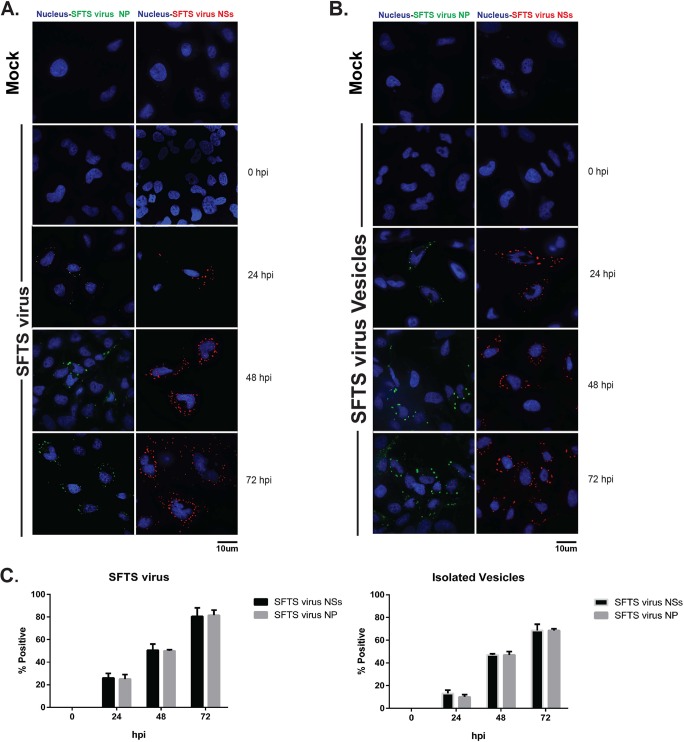
FIG 8.
Virions harbored within SFTS virus NSs-positive extracellular vesicles are capable of establishing productive infection. HeLa cells were mock infected (top of panels A and B), infected with SFTS virus (A), or infected with supernatants collected from cells infected with the purified, CD63 immune-selected extracellular vesicles described in Fig. 7D (B). Cells were fixed at 0, 24, 48, and 72 hpi. Immunofluorescence was performed using primary antibodies against SFTS virus NP or NSs and Alexa Fluor 488 as the secondary antibody. Nuclei were visualized with Hoechst 33342. Representative images for the mock-infected groups are shown. (C) Percentages of cells positive for SFTS virus NP or NSs calculated by cell counts in 10 fields of view. A total of 150 cells were counted, and the percentage was calculated by dividing the total cells positive for SFTS virus NP or NSs by 150.

