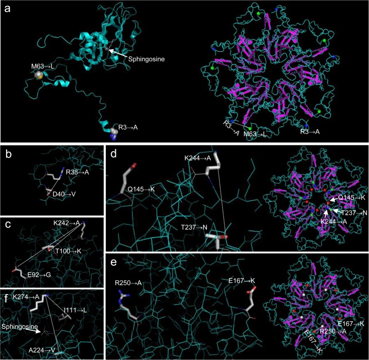
FIG 5.
Locations of mutants with second-site compensatory mutations in the structure model of EV71 VP1 (PDB 3VBS). The VP1 structure is shown in cyan, and for each side chain and the pocket factor sphingosine, elements are shown as follows: C, white; H, gray; N, blue; O, red; and S, orange. (a) Localization on a capsid protomer (left panel) and pentamer (right panel) of residues 3 (blue), where Arg was mutated to Ala, and 63 (green), where a second-site mutation from Met to Leu fully restored virus production. (b) Asp40 was mutated to Val to restore the infectivity of mutant R38A. (c) Glu92 and Thr100 were mutated to Gly and Lys, respectively, to restore the infectivity of mutant K242A. (d) Gln145 and Thr237 were mutated to Lys and Asn, respectively, to restore the infectivity of mutant K244A. The locations of residues Lys244 (blue), Gln145 (red), and Thr237 (green) are shown either on a VP1 monomer (left panel) or pentamer (right panel). (e) Localization on a capsid protomer (left panel) and pentamer (right panel) of residues 250 (white), where a lethal mutation from Arg to Ala was introduced, and 167 (red), where a second-site mutation from Glu to Lys partially restored virus production. (f) Localization on a capsid protomer of residue K274 and second-site mutation I111L and A224V, which partially restored the infectivity of lethal mutant K274A. This figure was produced using PyMol and Cn3D for protomer and pentamer, respectively.

