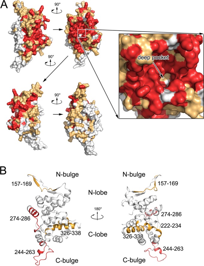
FIG 7.

Epitopes in hantavirus NPcore. (A) Sequence conservation mapping on the surface of hantavirus NPs. Primary sequence homologies of representative hantaviruses are defined, and the structure of SNV NPcore is shown in front, side, and back views, covering its molecular surface. The deep pocket inside the RNA-binding crevice is enlarged for detail. Strictly conserved residues are in red, while conserved and variable residues are in gold and white, respectively. (B) Epitopes related to serotype specificity in hantavirus NPcore. Previously reported epitopes on NPcore that can distinguish SNV and ANDV (64) are highlighted in the structure of SNV NPcore. The two regions with the most serotype specificity (i.e., 244 to 263 and 274 to 286) are in red; the other identified epitopes are in gold.
