
FIG 3.
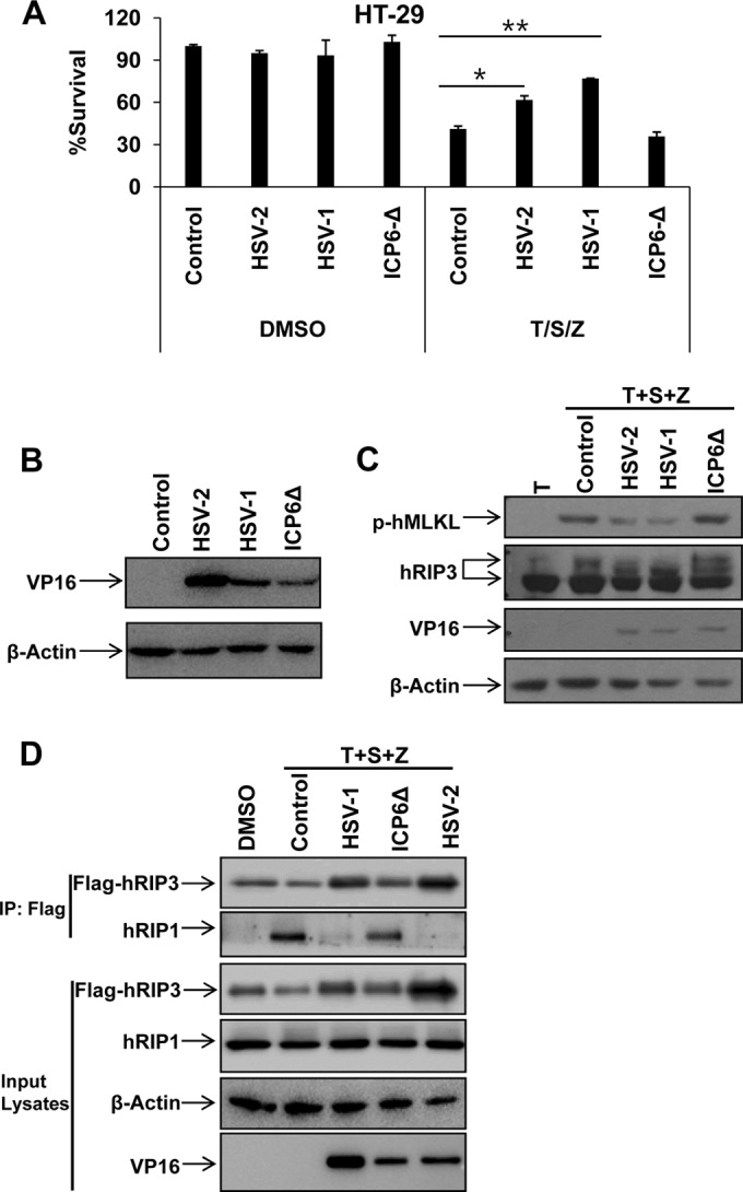
HSV R1 is required to disrupt necrosome formation during necroptosis in human cells. (A) HT-29 cells were infected with the indicated virus (MOI of 2.5). At 2 h postinfection, cells were treated with dimethyl sulfoxide (DMSO) or TNF-α/Smac mimetic/Z-VAD for 16 h, and then cell viability was determined. *, P < 0.05; **, P < 0.001 versus the control. (B) HT-29 cells were infected with the indicated virus (MOI of 2.5). Cell lysates were collected at 6 h postinfection and then subjected to Western blot analysis. (C) HT-29 cells were infected as indicated (MOI of 2.5). At 2 h postinfection, cells were treated with TNF-α/Smac mimetic/Z-VAD for an additional 6 h. Then cell lysates were collected and subjected to Western blot analysis. (D) HeLa-hRIP3 cells were infected as indicated (MOI of 2.5). At 2 h postinfection, cells were treated with TNF-α/Smac mimetic/Z-VAD for an additional 6 h. Cell lysates were collected and used for anti-Flag immunoprecipitation. The Flag-RIP3 immunocomplex was analyzed by Western blotting with the indicated antibody.