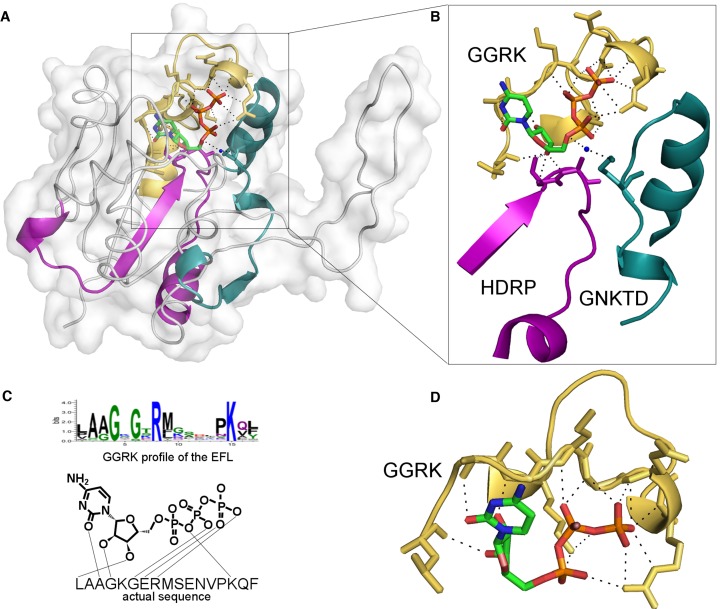
Figure 2.
Structure of 2-C-methyl-D-erythritol 4-phosphate cytidylyltransferase from Thermotoga maritima with bound CTP. UniProt accession number for the protein is Q9 × 1B3, PDB ID 1vpa. (A) Structure with three ligand-binding EFLs are displayed as colored ribbons. (B) Zoom-in to the CTP binding site. (C) Scheme of contact between sequence found by the ‘GGRL’ profile. (D) Structure of the motif (yellow) found by the ‘GGRK’ profile. This motif interacts with three parts of the ligand: base, ribose and phosphate groups. Motif found by the ‘HDRP’ profile interacts with ribose (magenta). Motif found by ‘GNKTD’ profile (green loop) makes a contact with phosphate groups via the water molecule.

