Fig. 5.
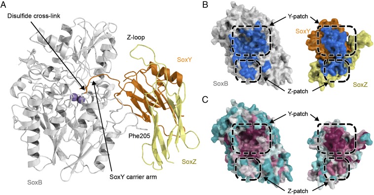
Structure of a disulfide-linked SoxB–SoxYZ complex. (A) Overall structure of the complex with proteins in cartoon representation and the manganese ions shown as purple spheres. Stick representation is used to show the disulfide bond between the SoxY carrier arm Cys and SoxB Cys175, and for SoxB residue Phe205 that contributes to the Z-patch. (B) Surface representation of the interacting faces of the SoxB and SoxYZ proteins. The surfaces that are buried upon interaction are shown in blue. (C) The same views of the SoxB and SoxYZ proteins as in B but with the surface colored according to sequence conservation using the program Consurf (22). Magenta indicates areas of highest sequence conservation and cyan the most variable sequences. Note that the Z-patch conservation in SoxB and SoxYZ is probably underreported because of alignment difficulties caused by insertions and deletions in adjacent sequences in the proteins from Purple Sulfur Bacteria and Green Sulfur Bacteria.

