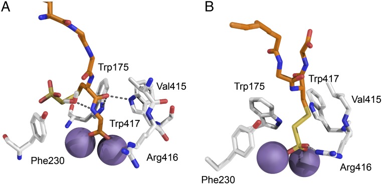
Fig. 9.
Models of the active site of the SoxB–SoxY(SSO3−)Z complex. Models were constructed based on the native SoxB–SoxYZ structure as described in SI Appendix, SI Materials and Methods. The carbon atoms of SoxB are shown in gray and of SoxY in orange. Oxygen atoms are red, nitrogen atoms blue, and sulfur atoms yellow. The manganese ions are represented by purple spheres. (A) Complex with the SoxY C-terminal carboxylate coordinating the SoxB manganese ions. The interaction of SoxB with the SoxY C-terminal Cys-Gly peptide is stabilized by hydrogen bonding. (B) Complex with the S-thiosulfonate group coordinating the manganese ions. The interaction of SoxB with the cysteine-S-thiosulfonate is stabilized by an arc of hydrophobic residues.

