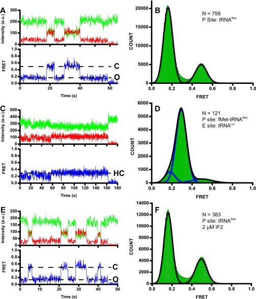
Fig. S8.
Observation of the L1 stalk in the open, closed, and half-closed positions using the L1/L33 FRET pair. Ribosomes preassembled from unlabeled 30S subunits and L1-Cy5/L33-Cy3 50S subunits contained a P-site deacylated tRNAfMet (A, B, E, and F) or a P-site fMet-tRNAfMet (C and D). In C and D, ribosomes were imaged in the presence of 100 nM tRNALys; in E and F, ribosomes were imaged in presence of 2 μM IF2 and 1 mM GDPCP. Representative smFRET traces (A, C, and E) show Cy3 and Cy5 fluorescence in green and red, respectively, and apparent FRET is shown in blue. Dashed lines, FRET values corresponding to closed (C), open (O), and half-closed (HC) states of the L1 stalk. Cy3 photobleaching is not shown for presentation purposes. FRET distribution histograms (B, D, and F) were fit the sum of two (B and F) or three (D) Gaussians. Individual Gaussian fits shown in blue with the sum of Gaussians shown as a black trace. N, number of traces used to assemble each histogram.

