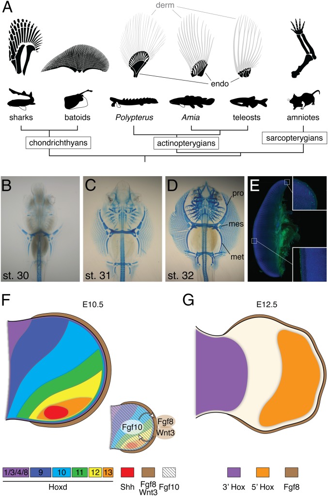
Fig. 1.
Appendage diversity and the skate unique fin. (A) The pectoral fin and forelimb skeleton in a variety of taxa. Chondrichthyan and basal actinopterygian fins are composed of three bones, pro-, meso-, and metapterygium, whereas the sarcopterygian appendage is a single rod of the metapterygium axis. The batoid fin is extremely wide along the A–P axis compared with other vertebrates. Dermal bones (derm) and endochondral bones (endo) are labeled by gray and black colors depending on the difference of its developmental mechanisms (36). (B–D) Alcian Blue-stained skeletal preparations of skate embryos at stages 30–32. mes, mesopterygium; met, metapterygium; pro, propterygium. Both pectoral and pelvic fins elongate along the A–P axis. (E) Immunostaining for phosphorylated histone H3 (green) and DAPI (blue) in the pectoral fin at stage 31. Image composed using tiled scanning by confocal microscope (Zeiss ZEN software). Inset, magnified portions of the anterior and central fin. Statistical analysis of cell proliferation rates in each portion can be found in SI Appendix, Fig. S1. (F and G) Summary of the developmental mechanisms of the tetrapod limb. At an early stage (F), Shh is expressed in the posterior limb bud, and 5′Hox genes show a gradient of expression. Fgf10 induces and maintains AER structure. In turn, Fgf8 and Wnt3 in AER stimulate cell proliferation in the limb mesenchyme. As the limb bud develops, 5′Hox genes mark the autopod domain, whereas 3′Hox genes are expressed in the proximal limb.

