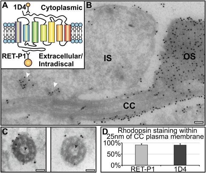
Fig. 1.
Rhodopsin is transported to the newly forming discs via the connecting cilium plasma membrane. Retinal sections were labeled with two rhodopsin antibodies: 1D4 (10 nm gold) against the cytoplasmic and RET-P1 (15 nm gold) against the extracellular/intradiscal epitope. (A) Rhodopsin diagram showing the 1D4 and RET-P1 antibody epitope sites. (B) Note accumulation of rhodopsin-bearing vesicles (white arrowheads) in the IS at the base of the connecting cilium (CC) and rhodopsin confined to the plasma membrane within the CC and the discs within OS. (C) Cross-sections of the CC showing rhodopsin staining restricted to the plasma membrane. Black arrowheads indicate small vesicles with sticklike projections within the CC. (D) Percentage of rhodopsin labeling present on the ciliary plasma membrane. (Scale bar, 100 nm.)

