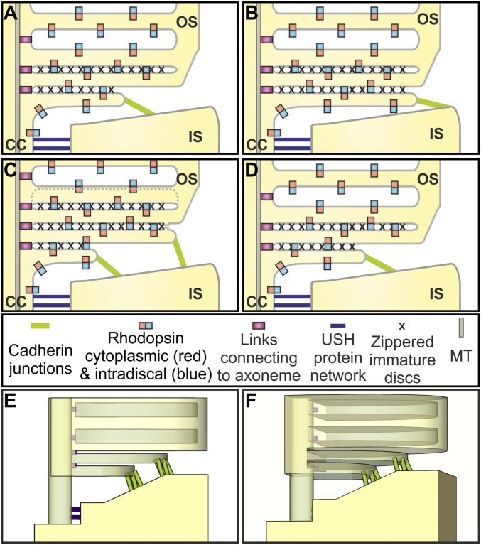
Fig. 7.
Model of maturation of rod photoreceptor discs. (A–D) Schematic showing the rhodopsin-containing plasma membrane of the connecting cilium that is linked to the side of the periciliary ridge by the USH protein network. The evaginating rhodopsin-containing membrane zippers up with neighboring evaginations proximally, leaving the leading edge free to interact with the top of the periciliary ridge via PCDH21-containing junctions. Discs at the base of the evaginations are anchored to the axoneme. The leading edges of neighboring evaginations fuse to form closed discs, accompanied by disassembly of the PCDH21-containing junctions and followed by change in membrane spacing to generate discs with a greater depth. (E and F) 3D models of the base of the OS showing cadherin junctions between developing discs and the periciliary ridge of the IS.

