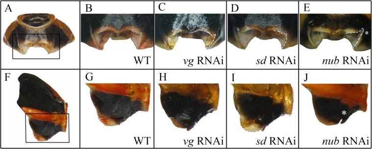
Fig. S3.
(A) Fronto-ventral view of T1 ventral plate. (B–E) close-up of region outlined in A in a wild-type, vg RNAi, sd RNAi, and nub RNAi, respectively. (F) The lateral view of T1 plate. (G–J) The magnified detail of the outlined region in wild-type (G), vg RNAi (H), sd RNAi (I), and nub RNAi (J), respectively. The asterisk in J shows a notch created in the nub RNAi treatment.

