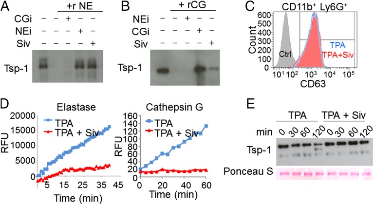
Fig. 4.
Neutrophil-derived Ser proteases mediate proteolysis of Tsp-1. (A and B) In vitro degradation assays of recombinant Tsp-1 with recombinant NE and CG proteases, alone or in combination with the specific inhibitors of NE or CG, and sivelestat. Recombinant (r) protein incubations were followed by Western blot analysis for Tsp-1 levels. Experiments were repeated three times with similar results. (C) Representative flow cytometry analysis of degranulation marker CD63 in CD11b+ Ly6G+ cells cultured in vitro with 0.01% DMSO (Ctrl, solid gray histogram), 20 nM TPA (TPA, empty blue histogram), or 20 nM TPA + 0.05 μg/μL sivelestat (Siv, solid red histogram). Experiments were repeated three times with similar results. (D) Representative protease activity assays for NE (Left) and CG (Right) in CM of Ly6G+ cells cultured in vitro with 20 nM TPA or 20 nM TPA + 0.05 μg/μL Siv. Activities are represented as RFUs as a function of time. Experiments were repeated twice with similar results. (E) Western blot analysis of Tsp-1 in CM of Ly6G+ cells cultured in the presence of TPA or TPA + Siv at t = 0, 30, 60, and 120 min. Ponceau S staining shows equal protein loading.

