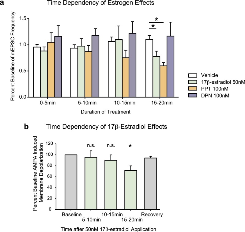
Fig. S8.
(A) Time dependency of rapid estrogen effects on mEPSC frequency. Vehicle group includes data from all three mEPSC experiments. Fifty nanomolars of 17β-estradiol and 100 nM of PPT decrease percent baseline mEPSC frequency only after 15–20 min of hormone treatment, but not at earlier time points. (Kruskal–Wallis H = 17.512, df = 3, P < 0.001, Dunn’s post hoc 50 nM 17β-estradiol: q = 2.396, df = 36, P < 0.05, 100 nM PPT: q = 3.944, df = 37, P < 0.05). (B) Time dependency of 17β-estradiol effects. This graph includes a combination of both experimental and pilot data examining estradiol effects on AMPA-elicited membrane depolarization amplitudes. Treatment of neurons with 50 nM of 17β-estradiol for greater than 15 min decreases AMPA neurotransmission. Significances indicate differences from baseline amplitudes using paired t tests (5–10 min: P = 0.9, 10–15 min: P = 0.8, 15–20 min: t = 2.54, df = 21, P < 0.05). Each treatment group represents an independent set of neurons, and the recovery measure is taken from a subset of neurons from the 15- to 20-min treatment group, following a 15-min washout with aCSF. *P < 0.05. n.s., nonsignificant.

