Fig. 3.
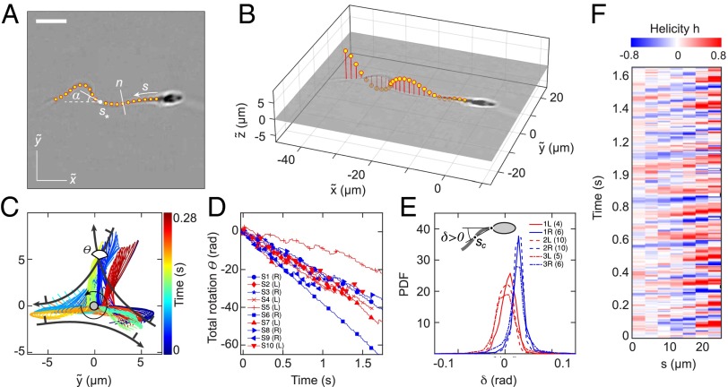
A 3D flagellar beat reconstruction reveals that a mirror symmetry breaking in the midpiece curvature separates left-turning from right-turning sperm. (A) A 2D bright-field image and tracked flagellum in the head-centered comoving frame, with arc-length s and normal line n. (Scale bar, 10 μm.) (B) A 3D beat reconstruction in the head-centered frame (Beat Reconstruction and Movie S7). (C) Typical 3D beat plane rotation for a single sperm, seen from head-on, with beat period of ∼0.08 s. The circular arrow indicates rolling direction of the flagellum. (D) The cumulative beat plane rotation θ, shown for 10 typical samples of left-turning (L) and right-turning (R) cells, implies that the rolling and turning direction are not correlated. (E) Midpiece curvature, quantified by the bend angle δ between the tangent at and the secant through μm, correlates strongly with the turning direction (three different donors, sample size in brackets). (F) A 3D reconstruction reveals ambidextrous helicity in the first ∼70% of the flagellum.

