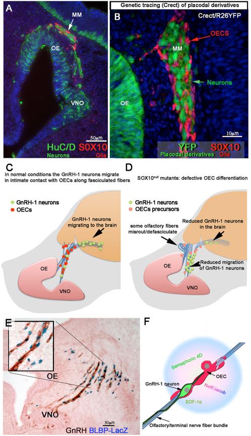
Fig. 7. The olfactory system is composed of placodal and neural crest cells. The OECs are important players in GnRH migration.
A) Section of mouse embryo E11.5 immunostained against HuCD (green, neurons) and Sox10 (Red, olfactory ensheathing cells (OECS)). Over development, the migratory mass (MM) composed of neurons and glia moves away from the olfactory (OE)/vomeronasal epithelia (VNO) toward the brain. B) Crect/RosaYFP genetic lineage tracing reveals placodal derivatives as positive for YFP expression (green). The OECs of the migratory mass are not derived from placodal progenitors. C and D) Diagrams (based on [150; 175]) representing normal GnRH neurons migrating along the olfactory/terminal nerve fibers from the nose to the brain in control animals; (C) In Sox10null mice, where the OECs do not complete maturation (pink dots), fewer GnRH neurons migrate into the brain and olfactory sensory axons misroute/defasisculate (D). Olfactory, vomeronasal and terminal fibers (blue), GnRH neurons green, OECs red in control, pink OECs precursors in Sox10null mutant. E) BLBP is expressed by the OECS; X-Gal staining (blue) on a section from BLBPCreIRESLacZ, [207], embryo, E12.5 immunostained for GnRH-1 (brown). GnRH-1 neurons migrate (brown) in close contact to maturing OECs (blue nuclei) (see insert). F) Model based on [127] representing OECS releasing molecules important for GnRH neuronal migration/motility on olfactory/terminal fibers.

