Figure 4.
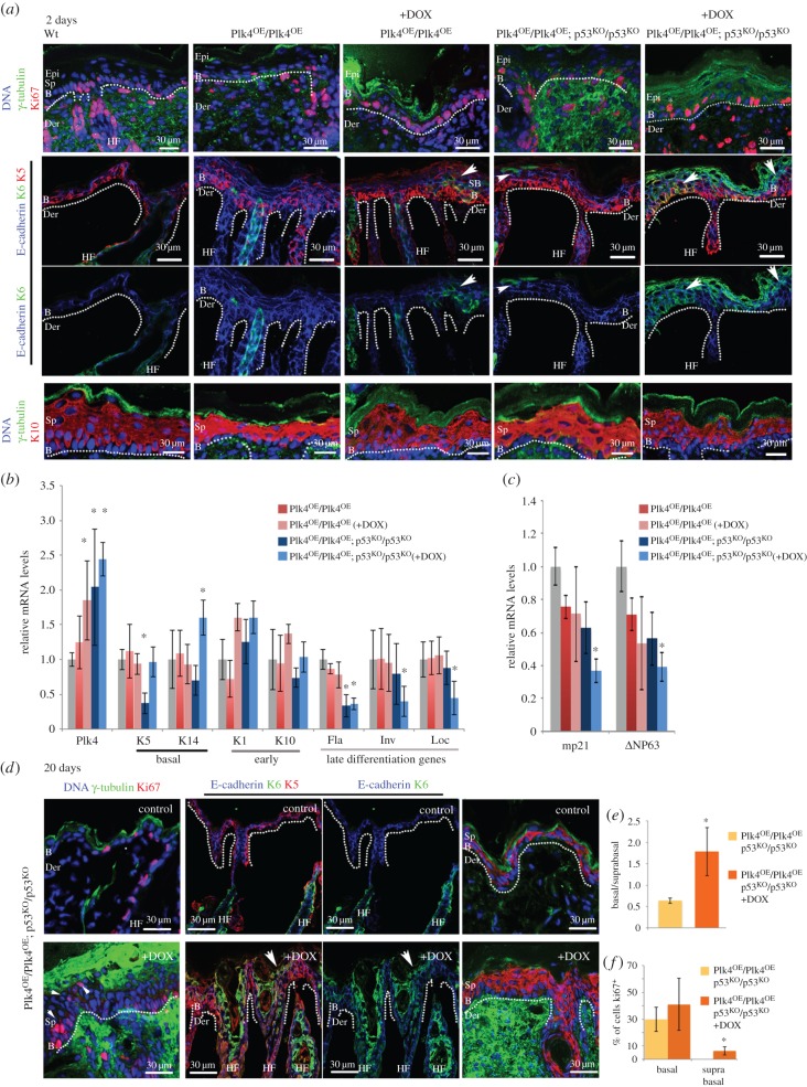
Plk4 over-expression leads to hyperproliferation and abnormal differentiation of cells in suprabasal layers. (a) Immunofluorescence of cryosections of P2 back skin from mice of indicated genotypes treated with doxycycline as indicated (+DOX). Note: Ki67 reveals proliferating cells in basal and suprabasal (white asterisk) epidermal layers of Plk4OE/Plk4OE; p53KO/p53KO (+DOX) mice; K6, restricted to hair follicles in controls, is present in epidermis following Plk4 induction (arrowheads). Basal layer marker K5 present throughout epidermis following Plk4 induction; distribution of early differentiation marker, K10 not affected and does not reveal differences between the experimental samples and the control at this stage. B, basal; Der, dermis; Epi, epidermis; Sp, suprabasal; dotted lines, dermo-epidermal border. (b) Quantitative RT-PCR assays for levels of indicated transcripts in total back skin from 2 day pups of indicated genotypes and treatment (n = 3 and three replicates for each sample). Note: elevated Plk4 transcripts consistently correlate with low expression of late differentiation genes filaggrin (Fla), involucrin (Inv) and loricrin (Loc). (c) Quantitative RT-PCR assays as in (b) for P21 (mp21) and ΔNP63. (d) Immunofluorescence analysis of back skin from 20 day pups of indicated genotypes and treatment. Note: Ki67-positive cells in basal and suprabasal cells (arrowheads) and K6 in multiple layers co-localizing with K5 (white arrow) in Plk4OE/Plk4OE; p53KO/p53KO (+DOX) mice; expanded staining of K5 and K10. (e) Ratio of basal to suprabasal cells indicated as mean ± s.d. (f) Proportion of cycling cells in epidermis. Mean ± s.d. % of cells showing Ki67-positive immunostaining. Significance determined by Student's t-test. *p < 0.05.

