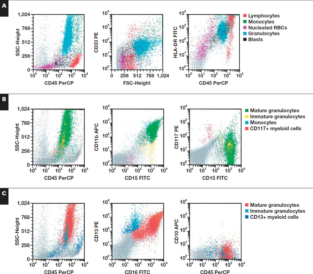
Image 3.
Representative flow cytometric findings in myeloid cells of a 21-year-old with Epstein-Barr virus–associated hemophagocytic lymphohistiocytosis (bone marrow). A, Granulocytes show normal SSC and appropriate expression of CD33 but are HLA-DR+. B, The granulocytes show appropriate expression of CD15 and CD11b and are CD117−. C, Myeloid cells show appropriate acquisition of CD13 and CD16 but express dim CD10. APC, allophycocyanin; FITC, fluorescein isothiocyanate; FSC, forward scatter; PE, phycoerythrin; PerCP, peridinin-chlorophyll protein; SSC, side scatter.

