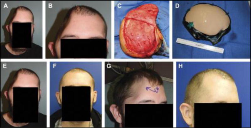
FIGURE 3.

Frontal view (A) and magnified view (B) of a hemicraniectomy patient at 1 month after infected bone flap removal. Right-sided bird’s eye view of pericranial onlay after careful dissection under loupe magnification and needlepoint electrocautery (C). Intraoperative preplating of a custom cranial implant by way of a sterile host bone model (D). Frontal view after bone flap removal (E) and the appearance at 2 months after reconstruction (F). Comparative right oblique views before and after reconstruction (G and H). Submental view demonstrates no signs of temporal hollowing (I).
