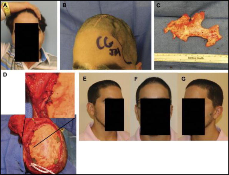
FIGURE 4.

Preoperative photographs at 1 year after autologous bone flap cranioplasty with severe resorption (A and B). Intraoperative photograph of thin, friable bone flap after removal with near-complete resorption (C). Secondary alloplastic cranial reconstruction required temporalis mobilization and fixation to the implant using a titanium plate and permanent suture fixation, allowing near-anatomical reconstruction (D). Right oblique, frontal, and left oblique views at 2 months showing acceptable contour and temporal symmetry (E–G).
