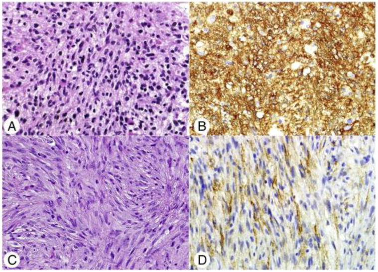
Fig. 4.

Histologic and immunohistochemical features of diffuse gliomas with NF1 loss. The histology of the 5 tumors with NF1 loss was variable, including fibrillary astrocytoma (H&E; original magnification ×400) (A) and 2 gliosarcomas The latter were characterized by neoplastic spindle cells (C), lacking GFAP expression and demonstrating pericellular reticulin staining (not shown). Matched podoplanin immunohistochemical stains demonstrating strong (B) and more modest (D) overexpression in these tumors (original magnification ×400).
