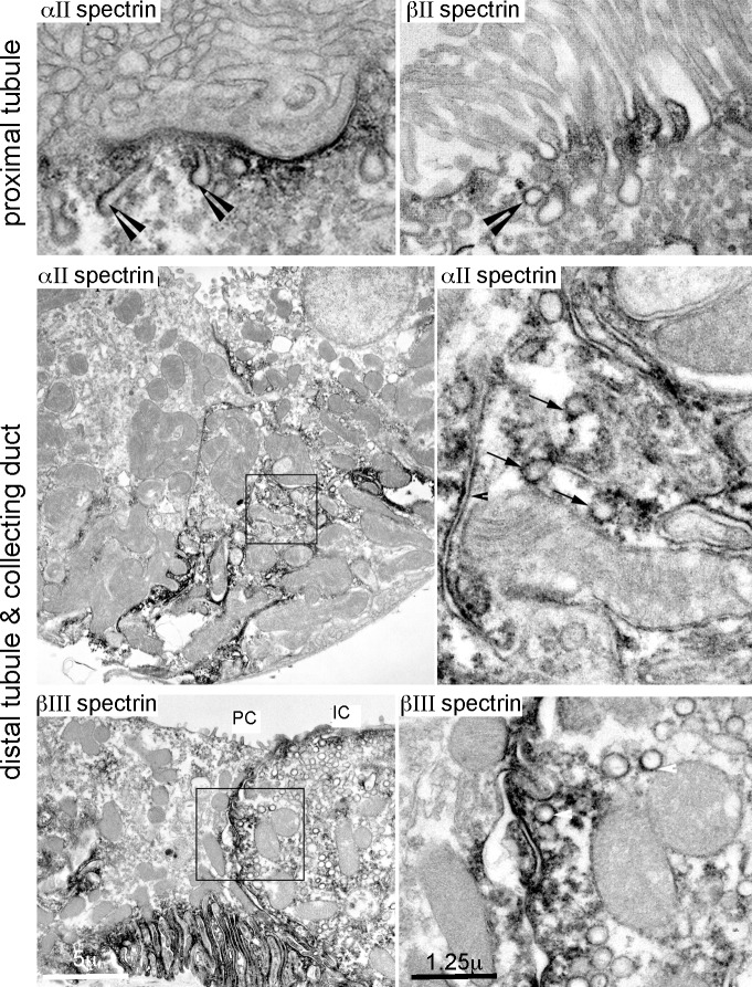
Fig 4. Spectrins associate with internal organelles.
ImmunoEM micrographs highlight a pool of αΙΙ/βΙΙ spectrin in association with a variety of organelles including canaliculi (arrow heads) and coated vesicles (arrows) in the cytoplasm and along the lateral and apical membranes of proximal and distal tubule cells and the collecting duct. The boxed areas are enlarged in the right column. PC, principal cell; IC, intercalated cell.

