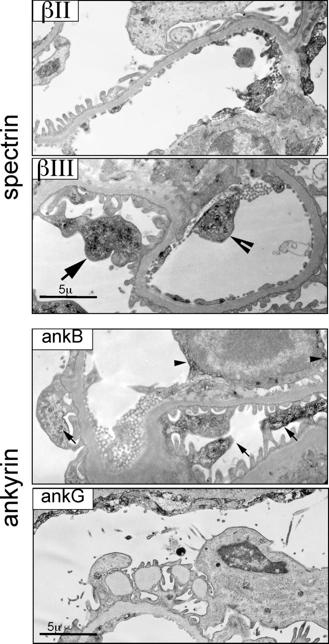
Fig 9. Spectrins and ankyrin distribution in the glomerulus.
ImmunoEM micrographs of spectrin and ankyrin in the glomerulus. Spectrin βΙΙ staining was largely confined to endothelial cells. Spectrin βΙΙΙ was present in both endothelial cells (arrow head) and podocytes (arrow). Ankyrin G largely spares the glomerulus except in the parietal layer of Bowman’s capsule. Ankyrin B was prominent in podocytes.

