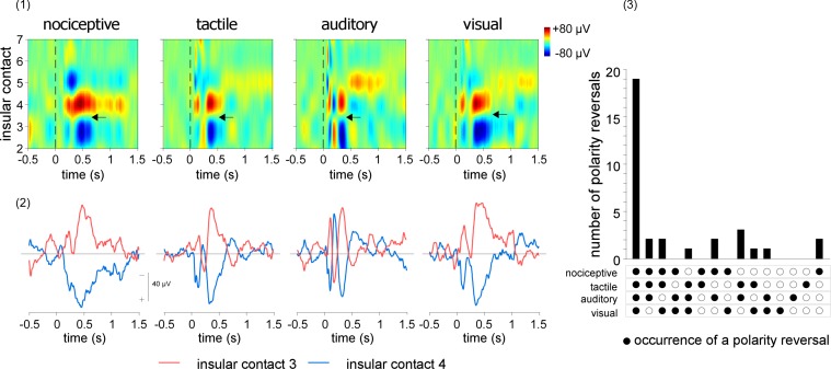
Fig 4. Linear CSD plots of the LFPs elicited by nociceptive, tactile, auditory, and visual stimuli delivered to the contralateral side.
Upper left panel. CSD maps obtained from the right insula of a representative patient, expressing the recorded signals as a function of time (x-axis) and insular electrode contact location (y-axis). Note that polarity reversals are observed at the same insular locations for all four types of LFPs. One of these polarity reversals is shown by the horizontal arrows, between contacts 3 and 4. Lower left panel. CSD signals recorded from these two contacts. The signal shown for each insular contact corresponds to the signal measured from that contact, using the average of the two adjacent contacts as reference. Right panel. Total number of polarity reversals that occurred at the same contact locations across modalities and patients. In almost all cases, polarity reversals occurred at the same sites for all four modalities, indicating that, at least at the mesoscopic level of intracerebral EEG recordings, the locations of the sources generating nociceptive and non-nociceptive LFPs in the insula are largely identical. doi:10.17605/OSF.IO/4R7PM.

