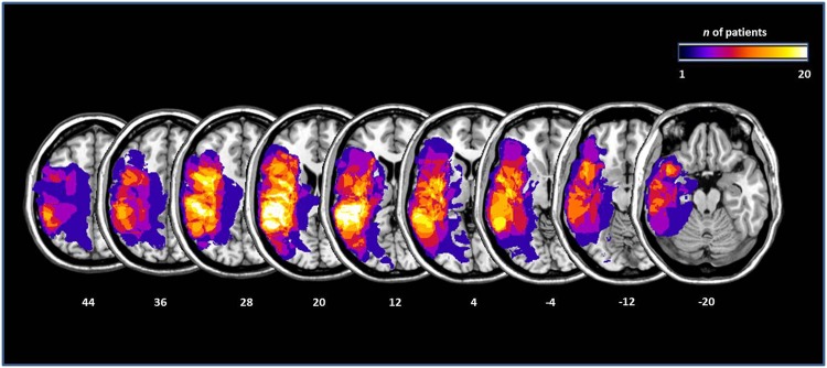
Fig 1. Lesions maps of the 20 aphasic patients, plotted on axial slices oriented according to the radiological convention.
Slices are depicted in 8mm descending steps. The Z position of each axial slice in the Talairach stereotaxic space is presented at the bottom of the figure. The number of patients with damage involving a specific region is color-coded according to the legend.

