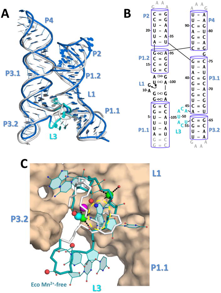
Figure 4. The Mn2+-free E. coli yybp-ykoY aptamer crystal structure.
A) Overall superposition of the E. coli Mn2+-free molecule B (blue) and L. lactis Mn2+-bound (gray) aptamer structures. The shifted L3 loop of the E. coli structure is in cyan. B) Secondary structure of the E. coli crystal structure, numbered according to wild-type sequence. C) Binding site close-up of the E. coli (teal) and L. lactis (gray) L3 loops. The L. lactis MA and MB are shown in cyan and bright pink. Two Sr2+ ions from the E. coli structure, one at MA and one in a new site, are shown in green. The phosphoryl oxygens in the E. coli Mn2+-free structure equivalent to those that bind MB in the L. lactis Mn2+-bound structure are shown as enlarged red spheres. The MA binding site is circled in yellow.

