Fig. 4.

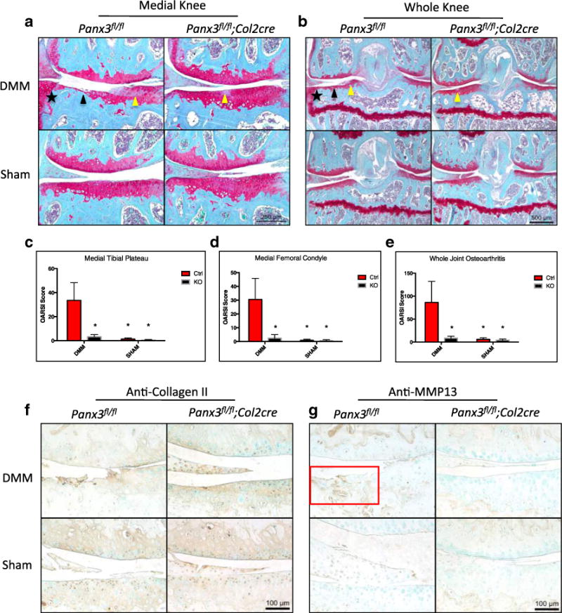
Chondrocyte specific loss of Panx3 protects against the development of surgically induced OA in mice. Representative images of Safranin-O/Fast Green-stained sections of Panx3fl/fl and Panx3fl/fl; Col2cre+/− DMM and Sham operated knees 8 weeks post-surgery (a, b). Black arrows indicate cartilage erosion. Yellow arrows indicate proteoglycan loss. Black stars indicate osteophyte formation. Medial knee and whole joint images were acquired. Cumulative OARSI scores of the medial tibia plateau (c), medial femoral condyle (d), and entire joint (e) are shown. Asterisk indicate means significantly different from Ctrl DMM scores (N=5, 4 DMM, sham per genotype; P<0.05, two-way ANOVA followed by Tukey’s test. Expressed as mean ± SEM. Serial sections from sham and DMM-operated joints harvested from Panx3f/f and Panx3f/f:Col2cre mice were immunostained with anti-collagen II and anti-MMP13 antibodies (f, g). Counter staining was done using methyl green. The red box highlights increased MMP13 immunolabeling within degenerated cartilage.
