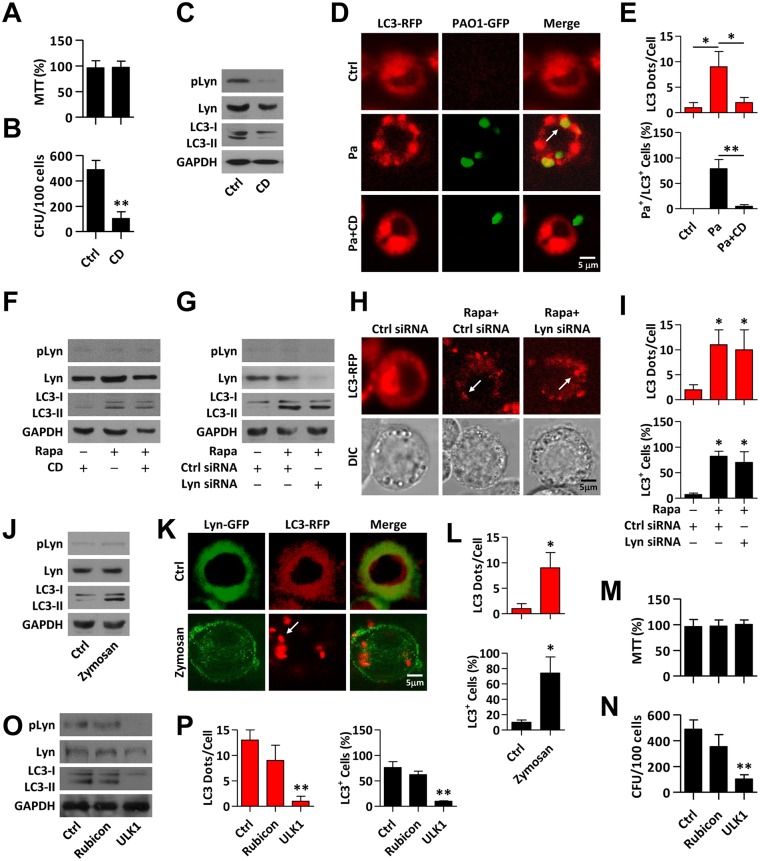
Fig 2. Lyn plays an important role in recruitment of LC3 upon Pa infection.
(A) MH-S cells were pretreated with CD (2.5 μg/ml, 30 min), and then infected with PAO1 (MOI = 10, 1 h). Cell viability was tested by MTT assay. (B) Cells treated as above were lysed for CFU assay. (C) Cells lysates were performed for immunoblotting of pLyn, Lyn and LC3. (D, E) MH-S cells were transfected with LC3-RFP plasmid for 24 h and pretreated with CD (2.5 μg/ml, 30 min). The cells were then infected with PAO1-GFP (MOI = 10, 1 h). Confocal microscopy images were used to detect LC3 puncta upon Pa infection. LC3 puncta in each cell were counted and the percentage of LC3+/Pa+ colocalization (LC3-RFP and PAO1-GFP) is shown. Data are derived from 100 cells in each sample. (F) MH-S were pretreated with CD (2.5 μg/ml, 30 min) or rapamycin (500 nM, 12 h) and cells were lysed for immunoblotting of pLyn, Lyn and LC3. (G) MH-S cells were transfected with Ctrl or Lyn siRNA for 24 h, and treated with rapamycin (500 nM, 12 h). Cells lysates were performed for immunoblotting of pLyn, Lyn and LC3. (H, I) MH-S cells were co-transfected with LC3-RFP and Ctrl or Lyn siRNA for 24 h. The cells were treated with rapamycin (500 nM, 12 h). Confocal microscopy images were used to show LC3 puncta. LC3 puncta in each cell were counted. The percentage of LC3+ events (cell with more than five LC3 puncta was considered as positive) is shown. Data are derived from 100 cells in each sample. (J) MH-S cells were treated with Zymosan (10 μg/ml, 1 h). Cell lysates were performed for immunoblotting of pLyn, Lyn and LC3. (K) MH-S cells were co-transfected with Lyn-GFP and LC3-RFP for 24 h. The cells were treated with Zymosan (10 μg/ml, 1 h). Confocal microscopy images were used to show Lyn or LC3 puncta. (L) LC3 puncta in each cell were counted, and the percentage of LC3+ events is shown as above. Data are derived from 100 cells in each sample. (M, N) MH-S cells were transfected with Ctrl, Rubicon or ULK1 siRNA for 24 h. The cells were infected with PAO1 (MOI = 10, 1 h). Cell viability and phagocytic abilities were tested by MTT or CFU assays as above. (O) Cell lysates from above were used for immunoblotting to detect pLyn, Lyn and LC3, respectively. (P) MH-S cells were co-transfected with Ctrl, Rubicon or ULK1 siRNA, respectively along with LC3-RFP for 24 h. The cells were infected with PAO1-GFP (MOI = 10, 1 h). LC3 puncta in each cell were counted, and the percentage of LC3+ events is shown as above. Data are derived from 100 cells in each sample. All data are representative as means+SD of three independent experiments. One-way ANOVA (Tukey’s post hoc); *, p<0.05; **, p<0.01. Scale bar = 5 μm.

