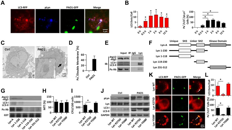
Fig 3. Lyn kinase activities are required to facilitate autophagy related phagocytosis.
(A) MH-S cells were transiently transfected with LC3-RFP. After 24 hours cells were infected with PAO1-GFP (MOI = 10, 1 h). Cells were fixed and stained with anti-pLyn antibody for immunofluorescence detection. Arrows indicate significant LC3 puncta colocalized with pLyn and Pa using CLSM imaging. Scale bar = 5 μm. (B) The statistic results from samples in A show LC3 puncta in each cell that are infected with Pa for different time. At 1 h post-infection, polymyxin B was added (1 h) to kill residual bacteria. The percentage of LC3+/Pa+/pLyn+ events (cell with colocalized puncta of LC3-RFP, PAO1-GFP, and pLyn) is shown. Data are derived from 100 cells in each sample. (C, D) MH-S cells were infected with PAO1 (MOI = 10, 1 h). After infection, cells were processed and examined by TEM. Arrow indicates autophagosome with double membranes containing internalized bacteria. The percentage of internalized Pa surrounded by double membrane is shown. Data are from 20 cells in each sample. Scale bar = 1 μm. (E) MH-S cells were infected with PAO1 as above. After infection, cell lysates were processed for co-IP to examine the interactions between Lyn and Atg12-Atg5, LC3 and Pa. (F) GST tagged Lyn peptide fragments were used to study in vitro association of Lyn with autophagy related proteins. (G) MH-S cells were infected with PAO1 (MOI = 10, 1 h) and then lysed for pull-down assay. GST-Lyn 1–230 containing both SH3 and SH2 domains shows association with Atg5-Atg12, LC3 and Pa. (H, I) MH-S cells were transfected with Lyn WT, DN (Lyn K275D), and constitutively active (Lyn Y598F) plasmid for 24 h and then treated with PAO1 as above. MTT assays were used to assess cell viability. CFU assays were used to measure phagocytosis. (J) Cells above were lysed for immunoblotting to detect the protein levels of pLyn, Lyn and LC3. (K, L) MH-S cells were transfected with Lyn WT, DN, and constitutively active plasmid for 24 h and then treated with PAO1-GFP (MOI = 10, 1 h). Confocal microscopy images were used to show significant LC3 puncta staining upon Pa infection. LC3 puncta in each cell were counted, and the percentage of LC3+/Pa+ events (cell with colocalized puncta of LC3-RFP and PAO1-GFP) is shown. Data are derived from 100 cells in each sample. Scale bar = 5 μm. All data are representative as means+SD of three independent experiments. One-way ANOVA (Tukey’s post hoc); *, p<0.05; **, p<0.01.

