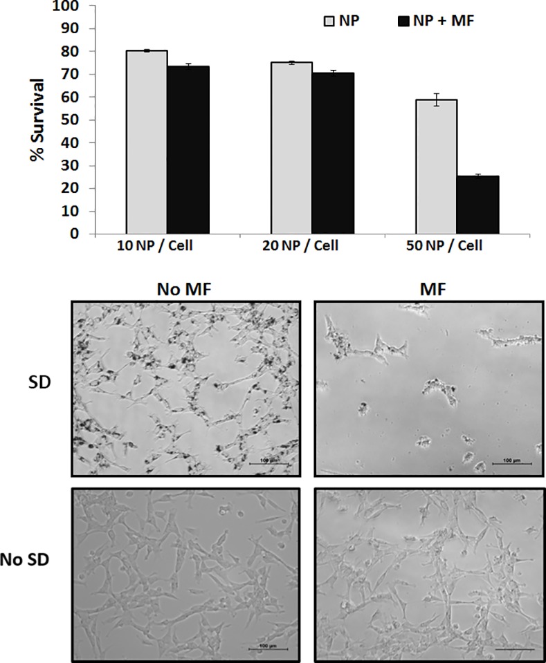
Fig 2. A: MTT assay of HB1.F3.CD cells. 10, 20, and 50 SD particles per cell were added into HB1.F3.CD cells for 24 hours. MF treatment consists of applying a 1T field rotating at 20 Hz for 30 min. B: Representative optical microscopy images of HB1.F3.CD cells before and after MF treatment.

The top panel contains SD and the bottom panels are control without SD.
