
Fig 1. Rheometry methods.
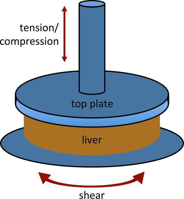
Liver samples were cut to a diameter of 20 mm and placed on a parallel plate rheometer with an upper platen of 25 mm. Tension and compression were generated by applying force in a direction perpendicular to the sample. Shear forces were applied by rotating the bottom plate in a direction parallel to the sample. Values derived from compression and tension studies were corrected to account for the difference in size between the sample and the top platen, and for narrowing at the waist of the sample in tension (see methods).