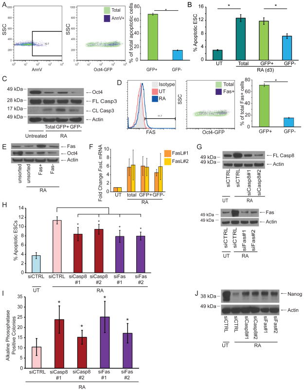
Figure 3. Fas and Casp8 Promote the Removal of Poorly Differentiating cells during ESC Differentiation.
(A) Representative FACS plots and quantification of Oct4-GFP ESCs stained for AnnexinV after 2d of RA treatment. (B) Quantification of apoptotic Oct4-GFP ESCs after 3d of RA treatment. (C) Immunoblots of lysates from 2d-treated Oct4-GFP ESCs after sorting for GFP expression. (D) Representative FACS plots and quantification of Oct4-GFP ESCs stained for Fas after 2d of RA treatment. RA-treated ESCs were used for isotype control staining. (E) Immunoblot for Oct4 from 2d-treated WT ESCs after sorting for Fas expression. (F) Q-PCR using two different primer sets for Fas-L mRNA from Oct4-GFP ESCs treated or untreated with RA for 2d. (G) Immunoblot of ESC lysates for Casp8 and Fas after siRNA knockdown and 2d RA treatment. (H) AnnexinV staining of ESCs quantified by FACS after indicated siRNA knockdown and 2d RA treatment. (I) Number AP+ colonies after indicated siRNA knockdown and 2d RA treatment. (J) Immunoblot of ESC lysates for Nanog after siRNA knockdown and 2d RA treatment. Three independent biological samples were used for the AnnexinV, qPCR, and AP assays. Data plotted as mean ± SD. * significant at p < 0.05 (Student t test).

