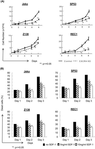
Figure 1.
(A) CXCR4 silencing in MCL cells leads to reduced cell growth. CXCR4 was silenced in four different MCL cell lines using a CXCR4 shRNA-encoding lentivirus or control lentivirus. After infection, cells were sorted for GFP and were selected using antibiotics. These stable cell lines (1×10⁴) were incubated in 24-well plates for 10 days to evaluate their growth. Cells were counted in triplicates every day.
(B) SDF-1 protects MCL cells from starvation-induced cell death. MCL cells (1×10⁶) were cultured in low serum for three days, and SDF-1 (5 ng/ml or 10 ng/ml) was added to the culture daily. The cells were evaluated using 7-AAD/Annexin V staining followed by FACS analyses.

