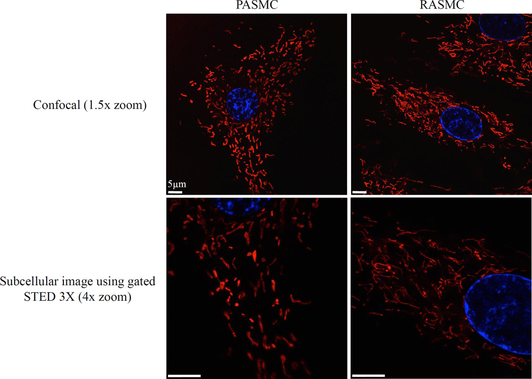
Fig. 2. Smooth muscle cell mitochondria are arranged in an extensive network.
Standard confocal (top) and super resolution (bottom) images of rat arterial smooth muscle cells. Cells stained with 50nM MitoTracker® Red FM and Hoechst 33342 (Molecular Probes, Life Technologies; Waltham, MA) for 20 min at 37°C and imaged using a Leica TCS STED 3X confocal microscope with and without stimulated emission depletion (STED); all images 100X oil with 1.5x or 4.0x zoom. A) Confocal image (1.5x) and B) STED confocal image (4.0x) of pulmonary artery smooth muscle cells. C) Confocal image (1.5x) and D) STED confocal image (4.0x) of renal artery smooth muscle cells

