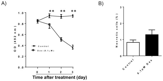
Figure 1. Effect of Dex treatment on INS-1 cell proliferation and viability.
A). INS-1 cells were cultured and treated with 0.1 μM Dex for different days. After treatment, cell proliferation rate was determined by MTT assay. B). After 48 hours of 0.1 μM Dex treatment, cell necrosis was determined by Trypan Blue staining. The percentage of necrotic cells was calculated. Data are expressed as mean ± SEM (n= 3 separate experiments). **, p<0.01 vs. 0 time point.

