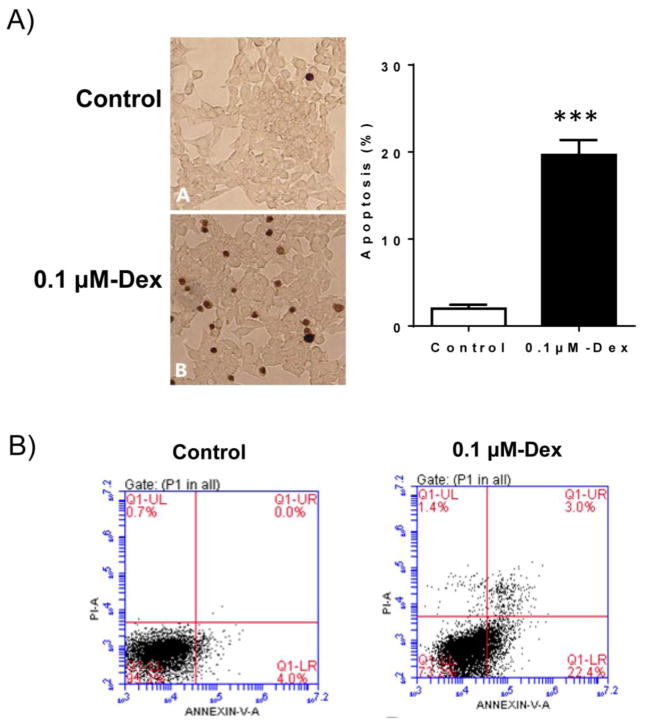
Figure 2. Dex treatment induced INS-1 cell apoptosis.
A). INS-1 cells were cultured and treated with 0.1 μM Dex for 48 hours. After treatment, cell apoptosis was determined by TUNEL staining and the representative light micrographs of TUNEL staining (brown nuclear) were shown. Percent of apoptotic cells was calculated. Data are expressed as mean ± SEM (n= 3 separate experiments). ***, p<0.001 vs. control.
B). Dex-induced apoptosis was also determined by flow cytometry (FACS) after Annexin V-FITC/PI staining of INS-1 cells. The representative FACS images were shown.

