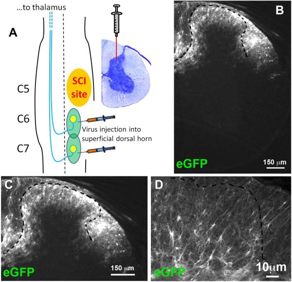
Figure 1. Intraspinal injections of AAV8-eGFP transduced superficial regions of the dorsal horn.
(A) Experimental paradigm showing unilateral C5 contusion and AAV8 injection into superficial dorsal horn at C6 and C7. (B) eGFP reporter expressing cells in dorsal horn. (C) Higher magnification image showing eGFP reporter expressing cells in dorsal horn. (D) Astrocyte-like morphology of eGFP+ cells.

