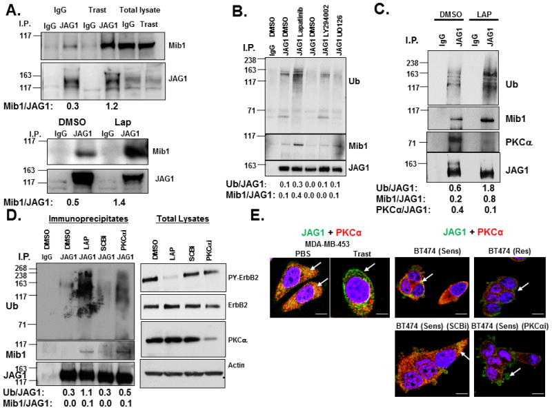
Figure 4.

ErbB-2 or PKCα Attenuates Mib-1-mediated Ubiquitinylation of Jagged-1. A, Trastuzumab or lapatinib increases the interaction between Mib-1 and Jagged-1. MDA-MB-453 cells were treated with controls (IgG or DMSO) or trastuzumab or lapatinib, respectively for 30 minutes. A Jagged-1 antibody was used to immunoprecipitate Jagged-1 from treated cell lysates. An IgG antibody was used as a control for nonspecific immunopreciptation. Western blotting was performed to detect co-immunoprecipitated proteins such as Mib-1 (Mib1). Jagged-1 (JAG1) protein was detected to determine specificity of the immunoprecipitation. Western blotting was also performed on total lysates to detect levels of total Mib1 and JAG1. B, Lapatinib-induced Jagged-1 ubiquitinylation is independent of PI3-K and MAPK signaling. Cells were treated with DMSO, lapatinib, LY294002, or U0126 and then lysed. Jagged-1 was immunoprecipitated and Western blotting was performed to detect ubiquitinylation (Ub), Mib-1, and JAG1. C, The recruitment of PKCα to Jagged-1 is inhibited by lapatinib. MDA-MB-453 cells were transfected with the FLAG-tagged Ub for 48 hours and treated with DMSO or lapatinib for 30 minutes. JAG1 was immunoprecipitated from cell lysates followed by western blotting to detect Ub, Mib1, PKCα, and JAG1. D, PKCα inhibits recruitment of Mib-1 and Jagged-1 ubiquitinylation. MDA-MB-453 cells were transfecteda a SCBi or PKCαi siRNA for 72 hours. Cells were then treated with DMSO or lapatinib for 30 minutes followed by Jagged-1 immunoprecipitation and western blotting to detect Ub, Mib1, and JAG1. Western blotting as was also performed on total lysates to detect PY-ErbB2, ErbB2, PKCα, and Actin. E, Jagged-1 and PKCα co-localize in ErbB-2 positive breast cancer cells. MDA-MB-453 cells were treated with PBS or trastuzumab for 48 hours. BT474 trastuzumab sensitive (Sens) and resistant (Res) cells were transfected with SCBi or PKCαi siRNA for 72 hours. Cells were fixed and stained for JAG1 (Green) and PKCα (Red) and visualized using Confocal immunofluorescence microscopy. The results are representative of three independent experiments. Left two panels scale bare = 10μm; right panels scale bars = 20μm. Image J software was used to measure densitometry of protein bands on Western blots. Ratios of proteins are presented below images.
