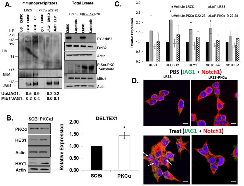
Figure 5.

PKCα is necessary and sufficient to inhibit Notch activation. A, Active PKCα prevents Mib1-mediated Jagged-1 ubiquitinylation. MDA-MB-453 cells were transduced with a retrovirus LRZS-linker or LRZS-PKCα 22–28 for 48 hours and then treated with DMSO or lapatinib for 30 minutes. Jagged-1 was immunoprecipitated from cell lysates followed by western blotting to detect Ub, Mib1, and JAG1 proteins. Total lysates were subjected with western blotting to detect total PY-ErbB-2, ErbB-2, P-Ser-PKC substrate, Mib1, and Actin. B, PKCα is necessary to inhibit Notch target gene expression. MDA-MB-453 cells were transfected with SCBi or PKCαi siRNA for 72 hours. Cell lysates were prepared and western blotting performed to detect PKCα, HES1, HEY1, and Actin proteins. Additionally, RNA was extracted from cells and real-time PCR was performed to detect relative expression of DELTEX1 transcripts normalized to HPRT. The results from the PCR represent means plus or minus standard deviation of three independent experiments. C, Active PKCα attenuates lapatinib-induced Notch target gene expression. MDA-MB-453 cells were transduced with a retrovirus LRZS-linker or LRZS-PKCα 22–28 for 48 hours and then treated with DMSO or lapatinib for 24 hours. RNA was extracted from cells and real-time PCR was performed to detect relative expression of HES1, DELTEX1, HEY1, NOTCH-1, and NOTCH-4 transcripts. The results are shown as means plus or minus standard deviations of three independent experiments. *Denotes statistical significance p<0.05 was calculated using an ANOVA. D, PKCα is sufficient to prevent trastuzumab-induced Notch-1 nuclear localization. BT474 trastuzumab sensitive cells were transduced with a retrovirus LRZS-linker or LRZS-PKCα for 48 hours and then treated with PBS or trastuzumab for 18 hours. Cells were fixed and stained for JAG1 (Green) and Notch-1 (Red) followed by visualization using Confocal immunofluorescence microscopy. Scale bars = 20μm.
