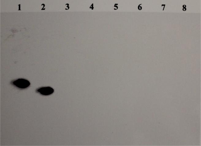
Fig. 6. RNase T1 protection assay.
Figure shows a chemiluminograph of 8M urea-6% polyacrylamide gel of RNase protection assay performed to detect apo II mRNA. Lane 1 shows the in vitro transcribed biotinylated antisense probe of upstream 3’-UTR of apo II mRNA that has irrelevant 5’ and 3’ overhangs, in the absence of RNase T1 digestion. Lanes 2-8, show RNase protection assays of whole cell RNA from livers of roosters treated with estrogen, resveratrol, genistein, catechin, tamoxifen, clomiphene and the vehicle, respectively. The band in lane 2 corresponds to the nuclease-protected biotinylated antisense probe of upstream 3’UTR of apo II mRNA.

