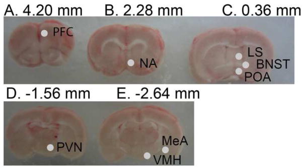
Figure 1.
Images of 5 brain sections from rostral to caudal (A–E) showing locations of 0.98 mm diameter punches. Punches are shown only on one hemisphere for ease in viewing, but bilateral punches were used for RNA extraction. Sections A–C are 2 mm thick, D–E are 1 mm thick. Abbreviations: Prefrontal cortex (PFC), nucleus accumbens (NA), lateral septum (LS), bed nucleus of the stria terminalis (BNST), preoptic area (POA), paraventricular nucleus (PVN), ventromedial hypothalamus (VMH), and medial amygdala (MeA).

