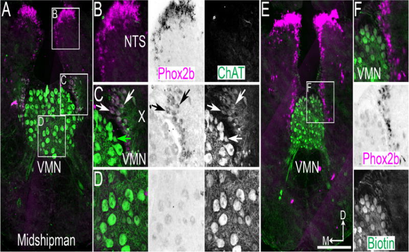
Figure 2.

Midshipman vocal motor nucleus (VMN) motoneurons do not express Phox2b. A. Confocal mosaic image of ChAT IHC (green) and pseudocolor inverted bright field ISH for Phox2b (magenta) in adult Midshipman caudal medulla. B–D. Expansions from A showing Phox2b and ChAT co-staining. Left – dual color image, middle – brightfield Phox2b staining, right – ChAT single color staining. Note the absence of ChAT staining in NTS region (B), co-localization of ChAT and Phox2b in vagal motoneurons (C), and absence of Phox2b expression above background in VMN (D). White arrows indicate co-expression, green arrow indicates ChAT only staining. E. Confocal mosaic image of fluorescent biotin labeling (green) and pseudocolor inverted brightfield ISH for Phox2b (magenta) in adult Midshipman caudal medulla. F. Expansion from E showing lack of biotin labeling in Phox2b expressing neurons. Top – color image, middle – brightfield Phox2b staining, bottom – fluorescent biotin labeling single color. Scale bar = 200 μm. D – dorsal, M - medial.
