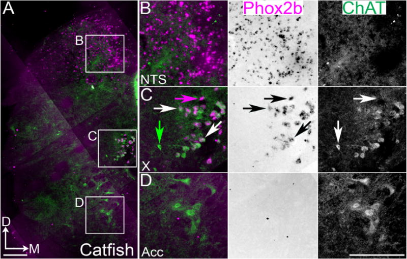
Figure 3.

Conserved organization of Phox2b populations in channel catfish caudal medulla. A. Confocal mosaic images of ChAT IHC (green) and inverted pseudocolor ISH for Phox2b (magenta) in adult channel catfish. B–D. Expansions from A showing combined (left) and single color images (middle – brightfield Phox2b, right – ChAT staining). B. Images showing Phox2b expression in non-cholinergic NTS neurons. Note the increased NTS size related to taste sensation. C. Images showing broad co-localization of Phox2b with ChAT in caudal vagal column. D. Images showing the absence of Phox2b mRNA expression in ChAT positive rostral spinal cord populations. White arrows indicate Phox2b and ChAT co-expression. Magenta arrow indicates Phox2b single expression. Green arrow indicates ChAT single expression. Scale bars = 200 μm. D – dorsal, M - medial. Acc – accessory nucleus.
