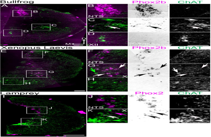
Figure 4.

Conserved organization of Phox2b populations in anuran and sea lamprey caudal medulla. Confocal mosaic images of ChAT IHC (green) and pseudocolor ISH for Phox2b (magenta) in premetamorphic bullfrog tadpole (A–D), or stage 55 African clawed frog (X. laevis) (E–H). B–D Expansions from A showing combined (left) and single color images (middle – brightfield Phox2b, right – ChAT) staining. B. Images showing Phox2b expression in non-cholinergic NTS neurons. C. Images showing broad co-localization of Phox2b with ChAT in the caudal vagal column. D. Images showing lack of Phox2b expression in XII motoneurons. F–H Expansions from E showing combined (left) and single color images (middle – Phox2b, left – ChAT) staining. F. Images showing Phox2b expression in non-cholinergic NTS neurons. G. Images showing broad co-localization of Phox2b with ChAT in caudal vagal column. H. Images showing lack of Phox2b expression in ventral motoneurons. I–K Confocal mosaic images of ChAT IHC (green) and pseudocolor ISH for Phox2 (magenta) in transformer sea lamprey. J–K Expansions from I showing combined (left) and single color images (middle brightfield Phox2, right ChAT) staining. J. Images showing Phox2b expression in non-cholinergic NTS neurons. K. Images showing co-localization of Phox2b with ChAT in presumptive vagal motoneurons. Note the presence of ChAT motoneurons lacking Phox2 expression ventral to vagal motoneurons in all species. White arrows indicate Phox2(b) and ChAT co-expression. Green arrows indicate ChAT single expression. Magenta arrow indicates Phox2 single expression. Note: X. laevis does not have a XII motor nucleus. Scale bars = 200 μm. D – dorsal, M - medial. NTS – nucleus, X – vagal motoneurons, XII – hypoglossal motoneurons.
