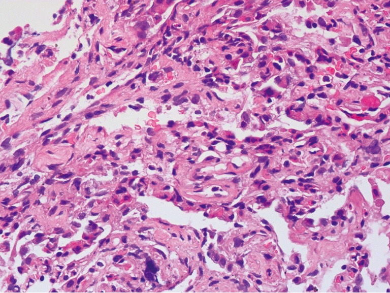
Figure 5.

The pathohistological findings from a limited post-mortem biopsy of lung tissue showed type II pneumocyte hyperplasia and shedding, diffuse pulmonary interstitial fibrosis, numerous lymphocytic infiltrates and only few neutrophilic infiltrates (haematoxylin & eosin staining, ×400).
