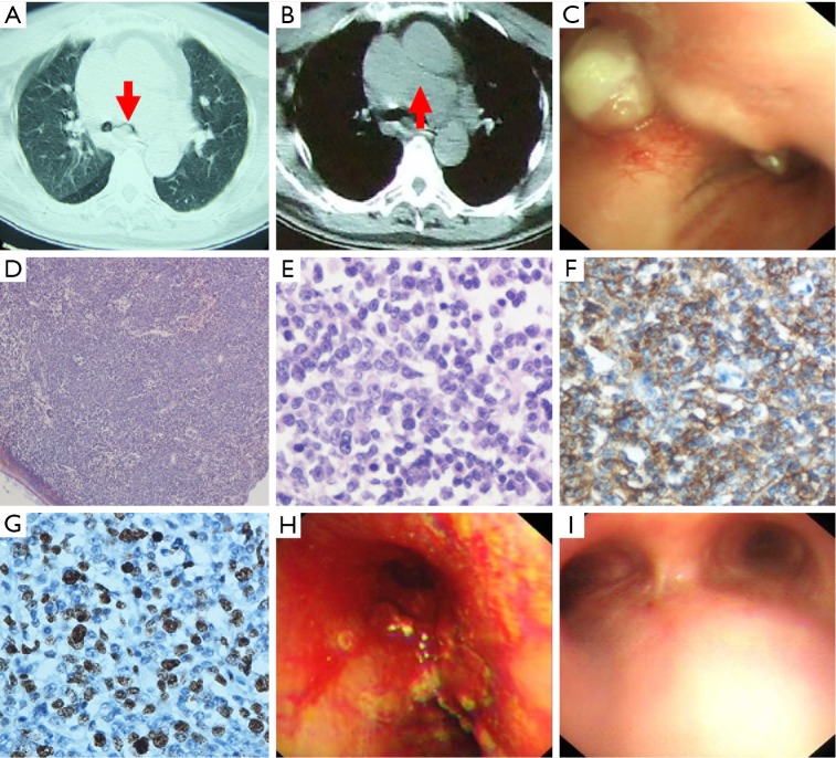
Figure 1.
The manifestation of chest computed tomography (CT), bronchoscopy and pathology. (A,B) CT of the chest on admission revealed a tumor blocking the left main bronchial stem (A, arrow shows the left bronchial stem), as well as enlarged lymph nodes compressing the trachea and main bronchus (B, arrow shows the enlarged lymph nodes); (C) bronchoscopic view of the trachea and carina showed widening of the carina. A large tumor was visible blocking the left main bronchial stem, and a smaller tumor was partially blocking the right main bronchial stem; (D-G) pathological examination of the tumor resected from the right bronchus showed immature lymphocytes; (H) after two interventional bronchoscopies, the main bronchi were rechanneled and some mucosal lesions remained; (I) bronchoscopic view after two cycles of R-CHOP chemotherapy showed complete recovery of the mucosa and tumor clearance from the airway; (D) hematoxylin-eosin staining revealed diffuse infiltration of atypical lymphoid cells with obvious dyskaryosis beneath intact bronchial mucosa (magnification, ×40); (E) hematoxylin-eosin staining revealed diffuse infiltration of atypical lymphoid cells with obvious dyskaryosis (magnification, ×400); (F) a diffuse infiltrate of large pleomorphic lymphoid cells stained positively for CD20, indicating high grade B-cell non-Hodgkin’s lymphoma (magnification, ×400); (G) the Ki67 proliferation fraction was 50% (magnification, ×400).

