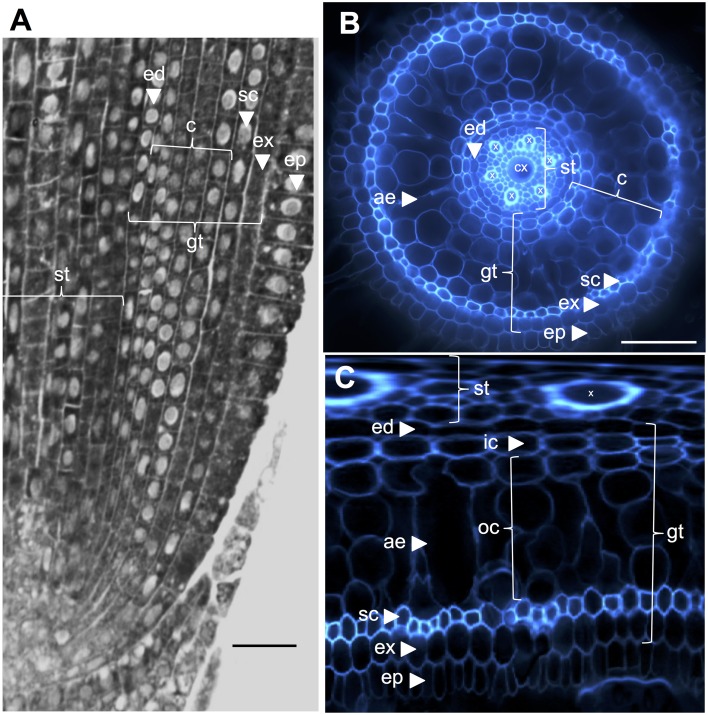
Figure 1.
Anatomy of a 6-day-old Nipponbare radicle. (A) Longitudinal section of a radicle fixed and paraplast embedded. Bar = 100 μm. (B) Transverse section of a fresh radicle observed under UV light. Bar = 50 μm. (C) Polar transformed view of B. Layers of tissues are labeled as follows: stele (st), ground tissue (gt) composed of several cell layers: one layer of endodermis (ed), several layers of cortex (c), one layer of sclerenchyma (sc), and one layer of exodermis (ex). The endodermal (ed) and exodermal (ex) layers are easily identified by the absence/reduction of fluorescence in the cell wall centers of these cells. The radicle is protected by one layer of epidermis (ep). Two types of cortical layers can be identified: inner cortex (ic), which is next to the endodermis (ed), and outer cortex (oc), which will eventually form aerenchyma (ae). In the stele, note the presence of a large central metaxylem (cx) surrounded by 6 metaxylem (x) vessels.

