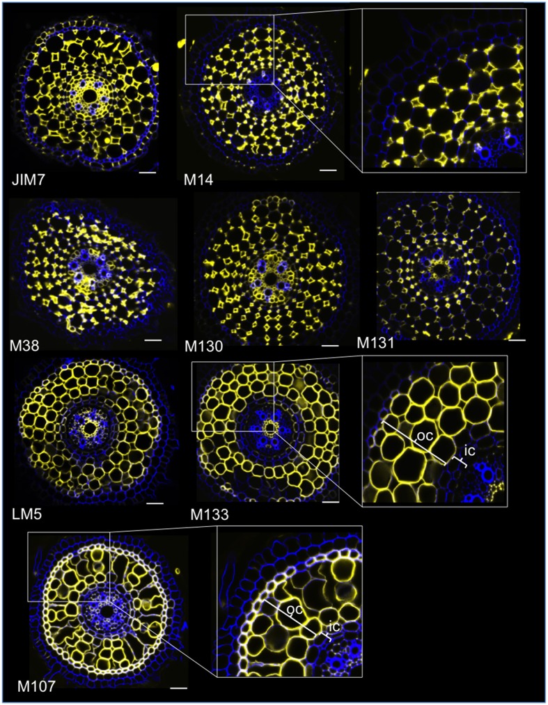
Figure 2.
Identification of eight specific cortex antibodies (JIM7, M14, M38, M130, M131, LM5, M133, and M107) among the 174 screened from the monoclonal cell wall antibody library. For each antibody, merged images of transverse sections were obtained under a confocal microscope after immunohistological labeling: in yellow, antibody pattern at 561 nm, and in blue, cell wall autofluorescence under UV light. Five antibodies (JIM7, M14, M38, M130, and M131) show a similar profile within all cortical cell layers stained (diamond shapes). Three antibodies (LM5, M133, and M107) homogeneously labeled cortical cell walls. Note that for these antibodies, inner cortical (ic) layers are not stained compared with outer cortical (oc) layers. Insets are close-ups of the corresponding images. Bars = 20 μm.

