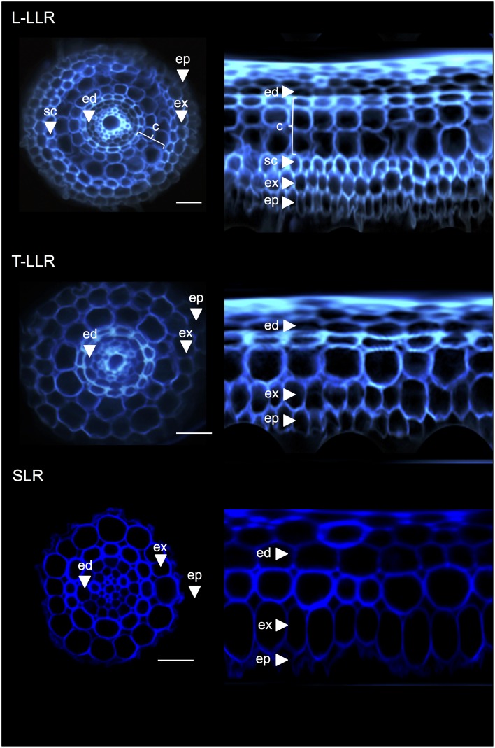
Figure 3.
Tissue composition of the Large Lateral Root types (Large-LLR and Thin-LLR) and Small Lateral Root (SLR). Seedlings were grown under hydroponic conditions for 2 weeks. Cell wall autofluorescence of transverse sections and polar transformed view under UV light. Tissues are labeled as follows: epidermis (ep), exodermis (ex), cortex (c), endodermis (ed). Note that the epidermis (ep) was often fragmented during the course of the experiment. Bars = 20 μm.

