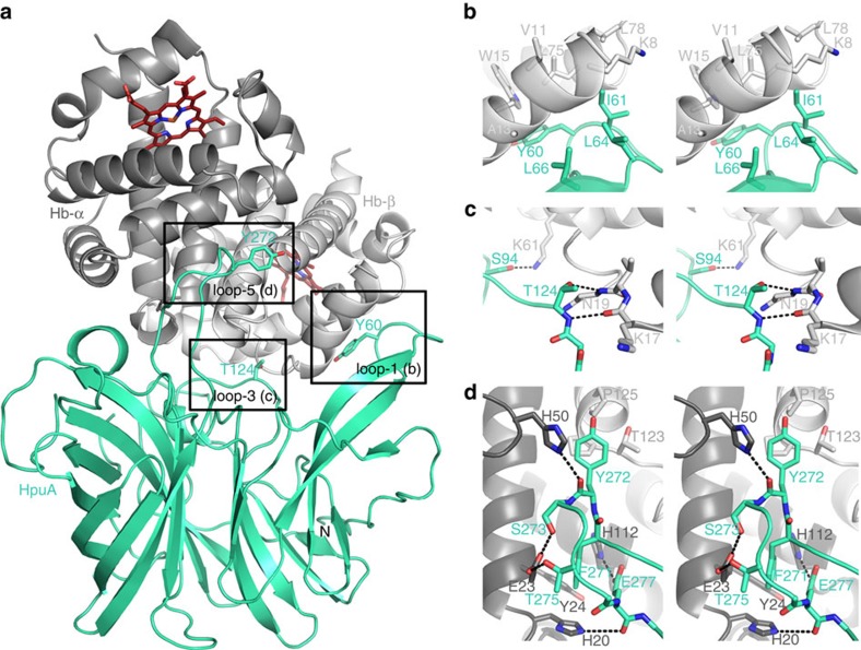
Figure 4. Crystal structure of the KdHpuA:Hb complex.
(a) Cartoon representation of half of the asymmetric unit, showing a KdHpuA monomer (cyan oriented as in Fig. 1b) binding to an α/β dimer of Hb (darker and lighter grey, respectively). The two haem groups of the Hb dimer (red) and key residues on four of the KdHpuA loops are shown as stick representations. (b) Close-up view in stereo of the interaction between KdHpuA loop-1 and Hb. Stick representations of important residues on both sides of the interface are shown. Carbon atoms are coloured as in a, oxygen atoms are red and nitrogen blue. (c) Stereo view of the interaction between KdHpuA loop-2 and loop-3, and Hb. Representations as in b with hydrogen bonds are shown as black dashed lines. (d) Stereo view of the interaction between KdHpuA loop-5 and Hb. Representation is conserved from a to c. A full list of interacting residues can be found in Supplementary Table 1.

